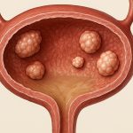
Bladder polypoid growths, ranging from benign papillomas to more concerning cancerous lesions, present a significant urological challenge. Traditionally, open surgery was the mainstay for removing larger or complex growths within the bladder. However, advancements in cystoscopic techniques have revolutionized management strategies, offering minimally invasive options that reduce morbidity and improve patient recovery. Cystoscopy allows direct visualization of the bladder interior, facilitating precise diagnosis and targeted treatment. This article will delve into the intricacies of excising large bladder polypoid growths utilizing cystoscopy, outlining the procedures, considerations, potential complications, and evolving techniques shaping this field. The goal is to provide a comprehensive overview for understanding how these growths are addressed with modern urological practices.
The shift towards cystoscopic excision isn’t merely about convenience; it’s about improved outcomes. Open surgery carries inherent risks associated with larger incisions, prolonged hospitalization, and extended recovery periods. Cystoscopy minimizes these drawbacks, allowing patients to return to normal activities sooner. Moreover, the precision afforded by cystoscopic techniques often results in better preservation of bladder function compared to more extensive surgical interventions. The decision-making process regarding whether to employ cystoscopic excision or pursue open surgery depends on several factors including growth size, location, characteristics (benign vs. malignant), patient health, and surgeon expertise – all carefully weighed to determine the optimal course of action for each individual case.
Cystoscopic Techniques for Polypoid Growth Excision
Several cystoscopic techniques are employed for excising bladder polypoid growths, chosen based on the growth’s size, location, and suspected pathology. Transurethral Resection of Bladder Tumor (TURBT) is arguably the most common approach. This involves using a resectoscope – a specialized instrument inserted through the urethra into the bladder – to visualize and then remove the growth in small pieces or as a single specimen. The resection relies on electrical energy, typically monopolar or bipolar, to cut and simultaneously cauterize tissue, minimizing bleeding. Holmium laser enucleation of the prostate (HoLEP) techniques have also been adapted for bladder polyp removal, offering precision and excellent hemostasis. More recently, en bloc resection using specialized instruments is gaining traction, aiming to remove larger growths as a single intact piece to aid in accurate pathological assessment and reduce the risk of tumor seeding.
The choice between monopolar and bipolar energy during TURBT impacts both efficacy and safety. Monopolar electrocautery uses a return pad placed on the patient’s thigh to complete the circuit, potentially causing complications like bowel perforation if not carefully managed. Bipolar electrocautery concentrates the current between two prongs of the instrument, minimizing stray currents and reducing the risk of such complications. Holmium laser enucleation offers advantages in terms of precision and hemostasis, making it particularly useful for larger or more vascular growths where bleeding is a concern. En bloc resection, while technically demanding, provides a more complete specimen for pathological analysis, crucial for accurate staging and grading, especially when malignancy is suspected. Understanding TURBT procedures can help patients better prepare for treatment.
Ultimately, the surgeon’s experience and familiarity with each technique play a critical role in choosing the most appropriate method. A thorough preoperative assessment including cystoscopy, imaging (CT or MRI), and potentially biopsy, guides this decision-making process. Postoperative follow-up is essential to monitor for recurrence and ensure complete eradication of the growth.
Considerations Before, During, and After Excision
Preoperative evaluation is paramount. This includes a detailed medical history, physical examination, urine cytology (to detect cancer cells), imaging studies (CT or MRI) to assess the extent of the growth and rule out invasion into the bladder wall or surrounding structures, and potentially blood tests to evaluate overall health and coagulation parameters. Patients should be informed about the procedure, its potential risks and benefits, and alternative treatment options. Preoperative bowel preparation may also be necessary depending on the technique employed. During the procedure, continuous monitoring of vital signs is essential, along with careful attention to fluid balance and bleeding control. The use of irrigation fluids during TURBT requires meticulous monitoring to prevent fluid overload or electrolyte imbalances – TUR syndrome – a potentially life-threatening complication.
Intraoperative considerations extend beyond technical aspects. Maintaining clear visualization throughout the resection is crucial for complete removal of the growth while minimizing trauma to surrounding healthy bladder tissue. Frequent biopsies should be taken from suspicious areas, even if they appear benign, as occult malignancy can sometimes be present. The en bloc approach requires careful dissection and a thorough understanding of the growth’s anatomy to avoid incomplete resection or injury to the bladder wall. Postoperatively, patients typically require a urinary catheter for several days to allow the bladder to heal and prevent bleeding. Close monitoring for hematuria (blood in the urine) is essential, as well as assessment for signs of infection or other complications.
Long-term follow-up is critical after cystoscopic excision. This includes regular cystoscopies with urine cytology to detect recurrence and monitor for disease progression. The frequency of follow-up depends on the initial pathology – benign growths require less frequent monitoring than malignant lesions. Patients who undergo resection for suspected bladder cancer will typically require more aggressive surveillance and potentially adjuvant therapies such as intravesical chemotherapy or immunotherapy to reduce the risk of recurrence. Early bladder cancer detection is crucial for successful treatment.
Managing Complications
Despite advancements in cystoscopic techniques, complications can occur. Hemorrhage is a relatively common complication, particularly with larger resections or when dealing with highly vascular growths. It’s usually managed conservatively with bladder irrigation and close monitoring; however, severe bleeding may require blood transfusion or even surgical intervention. Bladder perforation is another potential complication, though less frequent. Small perforations can often be managed conservatively with catheter drainage, while larger perforations may require surgical repair. Infection is also a risk, particularly after prolonged catheterization. Prompt diagnosis and treatment with appropriate antibiotics are essential to prevent sepsis.
A unique complication associated with TURBT is TUR syndrome, caused by absorption of large volumes of irrigation fluid into the systemic circulation. This can lead to hyponatremia (low sodium levels), hypothermia (low body temperature), and even cardiac arrest. Prevention involves using minimal amounts of irrigation fluid, monitoring serum sodium levels during the procedure, and promptly recognizing signs of fluid overload. Finally, urethral stricture – narrowing of the urethra – is a long-term complication that can occur after repeated cystoscopic procedures or due to scarring from thermal injury. It may require dilation or surgical correction.
The Role of Adjuvant Therapy
Adjuvant therapy plays a crucial role in preventing recurrence and improving outcomes for patients who undergo resection for suspected bladder cancer. Intravesical Bacillus Calmette-Guérin (BCG) is the gold standard adjuvant treatment for high-risk non-muscle invasive bladder cancer. BCG stimulates an immune response within the bladder, helping to eradicate residual tumor cells and prevent recurrence. However, it’s associated with side effects such as flu-like symptoms and cystitis. Intravesical chemotherapy, particularly mitomycin C, is another option for patients who cannot tolerate BCG or have failed BCG therapy.
The decision regarding which adjuvant therapy to use depends on the stage and grade of the initial tumor, the presence of high-risk features (e.g., carcinoma in situ), and the patient’s overall health. In some cases, systemic chemotherapy may be indicated for patients with more advanced disease or those who have progressed after intravesical therapies. Ongoing research is focused on developing new adjuvant strategies to improve outcomes and reduce recurrence rates in bladder cancer patients. Understanding specific types of bladder cancer informs treatment decisions.
Future Trends & Technological Advancements
The field of cystoscopic bladder polypoid growth excision continues to evolve rapidly. Robotic-assisted laparoscopy offers potential benefits in terms of precision, dexterity, and visualization for more complex resections or when dealing with large growths. Fluorescence guided surgery using agents like 5-aminolevulinic acid (ALA) helps delineate tumor margins, improving the completeness of resection. Advances in imaging techniques, such as narrow band imaging (NBI), enhance visualization of subtle mucosal changes, aiding in early detection and diagnosis.
Furthermore, research is focused on developing novel technologies for en bloc resection that minimize bladder wall trauma and improve specimen quality. Artificial intelligence (AI) and machine learning are being explored to assist surgeons with intraoperative decision-making, identifying suspicious areas and predicting recurrence risk. The ultimate goal is to develop more effective and less invasive approaches to manage bladder polypoid growths, improving patient outcomes and preserving bladder function for years to come. This evolving landscape promises a future where cystoscopic excision remains the preferred method for treating these challenging urological conditions. Robotic assistance can be beneficial in complex cases like posterior bladder wall mass resection.
A thorough preoperative assessment, including cystoscopy and imaging studies, guides the decision-making process. Consider exploring the purpose of a cystoscopy exam to understand its role in diagnosis.
Patients should also be aware of potential complications and the importance of postoperative follow-up.


