Kidney ultrasound and bladder imaging are both frequently utilized diagnostic tools in urology and nephrology, often employed to assess various conditions affecting these organs. While each technique provides valuable information independently, the question arises: can they be effectively combined? The answer is a resounding yes, and increasingly so with advancements in imaging technology. Combining these modalities not only streamlines the diagnostic process but also offers a more comprehensive understanding of urinary tract health – often within a single examination. This integrated approach allows clinicians to evaluate kidney structure and function alongside bladder characteristics like volume, wall thickness, and potential abnormalities, all without subjecting the patient to multiple separate procedures.
The synergy between kidney ultrasound and bladder imaging extends beyond mere convenience. A holistic assessment is crucial for accurate diagnosis because issues in one area often influence the other. For example, a blockage in the urinary tract can impact kidney function, while bladder dysfunction might contribute to kidney stone formation. Utilizing both modalities simultaneously provides a clearer picture of these interconnected relationships, leading to more informed treatment decisions and improved patient outcomes. Modern ultrasound machines are frequently equipped with capabilities that facilitate this combined imaging approach, making it a standard practice in many medical facilities.
Combining Ultrasound Modalities: Techniques & Benefits
The combination of kidney ultrasound and bladder imaging typically involves several techniques. A transabdominal ultrasound is the most common starting point for visualizing the kidneys – where a probe is placed on the abdomen to generate images using sound waves. To evaluate the bladder, the process often transitions into a suprapubic approach, focusing the ultrasound beam over the lower abdominal region where the bladder resides. However, transvaginal or rectal ultrasound can provide even more detailed views of the bladder in specific cases, particularly when assessing pelvic floor dysfunction or subtle abnormalities. Importantly, many modern machines allow for real-time switching between these approaches during a single exam.
The benefits of this combined approach are substantial. Firstly, it reduces patient discomfort and time commitment compared to undergoing separate ultrasound examinations or adding other imaging modalities like CT scans or MRIs. Secondly, the simultaneous evaluation allows for dynamic assessment – observing how the bladder fills and empties while simultaneously monitoring kidney function. This is particularly useful in diagnosing conditions like urinary retention, incomplete emptying, or vesicoureteral reflux (where urine flows backward from the bladder into the kidneys). Finally, integrated imaging improves diagnostic accuracy by providing a more complete picture of the entire urinary tract, aiding in identifying subtle connections between different symptoms and findings.
A key advantage is also cost-effectiveness. Ultrasound is generally less expensive than other advanced imaging techniques like CT or MRI, making combined kidney/bladder ultrasound an accessible initial step for many urological investigations. The information obtained can often dictate whether more complex – and costly – imaging is even necessary. This makes it a prudent choice in resource management within healthcare systems.
Assessing Hydronephrosis & Obstruction
Hydronephrosis, the swelling of a kidney due to a blockage, is frequently investigated using combined ultrasound techniques. When performing a kidney ultrasound, clinicians can quickly identify dilated renal collecting systems – indicating urine buildup. Simultaneously imaging the bladder helps determine if this dilation is caused by an obstruction within the urinary tract itself (like a kidney stone or tumor) or related to bladder dysfunction. If a blockage is suspected, Doppler ultrasound can be used to assess blood flow within the kidneys, helping differentiate between acute and chronic hydronephrosis.
The process often involves assessing the ureters – the tubes connecting the kidneys to the bladder. While ultrasound doesn’t always visualize the entire length of the ureter clearly, it can detect dilated ureteral segments or identify potential points of obstruction. When combined with bladder imaging, clinicians can evaluate the bladder wall thickness and look for signs of increased pressure caused by backflow from the obstructed kidney. This integrated assessment is crucial for determining the severity of the hydronephrosis and guiding appropriate treatment decisions, ranging from conservative management to surgical intervention.
Furthermore, post-operative evaluation benefits greatly from this combined approach. Following a procedure like ureteroscopy or stent placement, ultrasound can monitor kidney function and assess for any complications such as residual obstruction or infection. The ability to visualize both the kidney and bladder simultaneously allows clinicians to track healing progress and adjust treatment plans accordingly.
Evaluating Bladder Wall Abnormalities & Masses
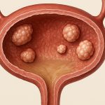
Ultrasound is excellent at characterizing bladder wall abnormalities. Combined with kidney imaging, it provides a more comprehensive assessment of potential causes. For example, identifying a mass within the bladder requires differentiating between benign growths (like polyps) and cancerous tumors. Ultrasound can assess the size, shape, and echogenicity (how sound waves reflect off the mass), providing initial clues about its nature. However, evaluating kidney function alongside this finding is vital – as kidney issues can sometimes mimic bladder abnormalities or influence their development.
When assessing a suspected bladder tumor, clinicians often use Doppler ultrasound to evaluate blood flow within the mass. Increased vascularity (more blood vessels) typically suggests malignancy. Simultaneously, they will examine the kidneys for any signs of metastatic disease or secondary effects from a potential primary bladder cancer. This holistic approach helps determine the extent of the disease and guide further investigations, such as cystoscopy (a direct visual examination of the bladder).
It’s important to remember that ultrasound is often used as an initial screening tool. If a suspicious mass is identified, more definitive diagnostic tests – like biopsy or CT/MRI – are usually required for confirmation. However, combined kidney and bladder ultrasound provides valuable preliminary information and helps streamline the diagnostic process.
Assessing Urinary Retention & Incomplete Emptying
Urinary retention (the inability to completely empty the bladder) can have significant consequences for both bladder and kidney health. Combined ultrasound imaging plays a critical role in diagnosing this condition. Post-void residual volume (PVR) – the amount of urine remaining in the bladder after urination – is easily measured using ultrasound. A high PVR suggests incomplete emptying, which can lead to urinary tract infections, hydronephrosis, and even kidney damage over time.
During the evaluation, clinicians will assess both the kidneys and bladder before and after voiding. This allows them to identify any underlying causes of retention, such as an enlarged prostate (in men), pelvic floor dysfunction, or neurological conditions. The simultaneous visualization of the kidneys helps determine if chronic retention has led to hydronephrosis – indicating a potential need for immediate intervention. Doppler ultrasound can also assess blood flow within the bladder wall, providing clues about its functional capacity and identifying any signs of chronic overdistension.
The combination is particularly useful in monitoring treatment effectiveness. Following interventions like catheterization or medication for prostate enlargement, ultrasound can track PVR and assess kidney function to ensure that drainage has improved and hydronephrosis has resolved. This iterative assessment ensures optimal patient care and prevents long-term complications associated with urinary retention.


