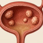
Cystoscopy is a cornerstone diagnostic procedure in urology, playing a vital role in evaluating a wide range of bladder conditions. It’s more than just looking inside the bladder; it’s about gaining crucial information to guide treatment decisions and improve patient outcomes. From investigating unexplained hematuria (blood in the urine) to monitoring response to cancer therapies, cystoscopy provides direct visualization that other imaging modalities simply can’t match. A thorough understanding of this procedure – what it entails, how it’s performed, and what findings might indicate – is essential for both healthcare professionals and patients alike navigating potential bladder health concerns.
The ability to directly visualize the bladder lining allows clinicians to identify subtle abnormalities that could be missed on scans or through symptoms alone. While imaging techniques like CT scans and MRIs offer valuable external information about the bladder’s size, shape, and surrounding structures, they lack the granular detail needed to assess the mucosal surface – where many lesions originate. Cystoscopy allows for targeted biopsies, ensuring accurate diagnoses and personalized treatment plans. It’s a procedure that bridges the gap between suspicion and certainty, providing essential clarity in complex cases.
Understanding the Cystoscopic Evaluation Process
Cystoscopy involves inserting a thin, flexible or rigid tube with a camera attached (the cystoscope) into the urethra and advancing it into the bladder. It’s typically performed as an outpatient procedure, though circumstances may necessitate an inpatient stay. Before the procedure, patients are generally given detailed instructions regarding bowel preparation and hydration to optimize visualization. Local, regional, or general anesthesia is used depending on patient preference, complexity of the expected findings, and available resources. A flexible cystoscopy is often preferred for initial evaluation due to its greater comfort and ease of use, while rigid cystoscopes provide superior optics and are typically employed when biopsies or interventions are needed.
The procedure itself usually takes between 15-30 minutes. During the examination, the bladder is filled with sterile fluid – commonly saline – to expand it and allow for a comprehensive view of the entire lining. The physician carefully inspects the bladder wall, looking for any irregularities such as tumors, inflammation, ulcers, or stones. Any suspicious areas are documented meticulously, often with photographic evidence, and biopsies can be taken at the time of the procedure. Post-cystoscopy, patients may experience some mild discomfort, burning during urination, or a small amount of blood in their urine – these symptoms usually resolve within 24-48 hours.
Crucially, cystoscopy isn’t just about finding problems; it’s also about ruling them out and providing reassurance to patients experiencing concerning symptoms. It helps differentiate between benign and malignant lesions, guiding appropriate follow-up care. The quality of the examination depends heavily on proper bladder preparation, a skilled operator, and adequate visualization achieved through sufficient fluid distension. Understanding potential causes for a thickened bladder wall with focal tumors is also key to accurate diagnosis.
Recognizing Bladder Wall Lesions Through Cystoscopy
The appearance of bladder wall lesions during cystoscopy is highly variable and dependent on the underlying pathology. Tumors often present as raised or ulcerated areas, though they can sometimes be flat and difficult to detect without careful scrutiny. Inflammatory conditions like cystitis may cause redness, swelling, and granular changes to the bladder lining. Stones appear as distinct shadows on the cystoscopic view, while diverticula (small pouches) are recognized as outpouchings from the main bladder wall.
Distinguishing between benign and malignant lesions solely based on visual appearance can be challenging. This is where biopsies become indispensable. Biopsies are usually taken using small forceps passed through the cystoscope, targeting any suspicious areas identified during the evaluation. These samples are then sent to a pathologist for microscopic examination to determine the nature of the lesion and guide subsequent treatment decisions. The ability to perform transurethral resection of bladder tumor (TURBT) simultaneously with diagnostic cystoscopy is a significant advantage, allowing for immediate removal of cancerous tissue in many cases. Patients may also benefit from understanding immunotherapy options for bladder cancer.
Important: Cystoscopic findings are always interpreted in conjunction with other clinical information, including the patient’s medical history, symptoms, and results from other tests. A comprehensive evaluation ensures accurate diagnosis and personalized treatment planning.
Biopsy Techniques & Interpretation
Biopsies during cystoscopy aren’t a one-size-fits-all process. The technique used depends on the lesion’s characteristics and location. – Forceps biopsies are commonly employed for smaller, easily accessible lesions. They involve using small grasping instruments to collect tissue samples. – Cold cup biopsies utilize a suction device to capture larger areas of suspicious mucosa. This is useful for evaluating diffuse or flat lesions. – Transurethral resection, as mentioned earlier, can serve both diagnostic and therapeutic purposes by removing the entire lesion for pathological analysis.
Following biopsy collection, the tissue samples undergo histological examination – meaning they are processed and examined under a microscope by a pathologist. The pathologist’s report will detail the type of cells present, their grade (indicating how aggressive the cells appear), and stage (describing the extent of disease). The report provides crucial information for determining the appropriate course of action, ranging from close observation to surgical intervention or chemotherapy. Accurate interpretation by both the urologist and pathologist is paramount for optimal patient care.
The Role of Image Enhancement Technologies
Traditional cystoscopy relies on white light illumination, which can sometimes make it difficult to identify subtle lesions. Fortunately, advancements in image enhancement technologies are improving diagnostic accuracy. – Blue light cystoscopy (BLC) utilizes a special filter that causes cancerous tissue to appear bright blue against a pale pink background, enhancing visualization of early-stage tumors. This is particularly useful for detecting carcinoma in situ (CIS), a non-invasive form of bladder cancer. – Narrow band imaging (NBI) enhances the contrast between normal and abnormal tissues by selectively filtering light wavelengths. This allows clinicians to better visualize vascular patterns and mucosal surface irregularities.
These technologies don’t replace the need for biopsies, but they significantly improve lesion detection rates and guide biopsy targeting. They allow for more precise assessment of the bladder lining and can potentially reduce the risk of missing early-stage cancers. The integration of these technologies into routine cystoscopic practice is becoming increasingly common as healthcare providers strive to provide the most accurate and comprehensive diagnoses possible.
Managing Patient Anxiety & Expectations
Undergoing a cystoscopy can understandably cause anxiety for many patients. Clear communication and patient education are essential components of pre-procedural care. Patients should be informed about what to expect during the procedure, including potential discomfort and post-operative symptoms. Addressing their concerns and answering their questions thoroughly can significantly reduce anxiety levels. Honest and empathetic communication builds trust and facilitates a positive patient experience.
It’s also important to manage expectations regarding the results of the cystoscopy. Patients should understand that biopsies may be necessary, and it could take several days or weeks to receive the final pathology report. The urologist should explain the potential implications of different biopsy findings and outline the next steps in the evaluation process. Providing support and reassurance throughout the entire experience is crucial for helping patients navigate this potentially stressful time.


