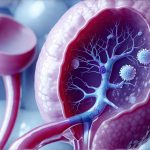
Interstitial nephritis (IN) represents inflammation within the kidney’s interstitial space – essentially the areas between the kidney tubules. It’s not a primary disease in itself as much as it is often a secondary consequence of something else, like an infection, medication side effect, or autoimmune disorder. While many associate kidney problems solely with glomerular diseases (affecting the filtering units), IN can significantly impact overall kidney function and potentially lead to chronic kidney disease if left unaddressed. Understanding its nuances is crucial because it sometimes presents subtly, making early diagnosis challenging. It’s vital to remember that kidney health is often interconnected; a problem in one area can ripple through others, influencing seemingly unrelated issues like stone formation.
The connection between interstitial nephritis and kidney stones isn’t immediately obvious to many people. Typically, we think of dehydration or high oxalate levels as the main culprits behind kidney stone development. However, IN disrupts the delicate balance within the kidneys, altering their ability to regulate various substances, including those that contribute to stone formation. This disruption can create a favorable environment for crystallization and subsequent stone development. Furthermore, some causes of interstitial nephritis themselves – certain medications or underlying conditions – can independently increase stone risk, creating a complex interplay between disease and consequence. This article explores the ways in which interstitial nephritis might raise the risk of kidney stones, examining mechanisms, related factors, and potential preventative strategies.
The Interplay Between Inflammation & Stone Formation
Interstitial nephritis fundamentally changes the microenvironment within the kidney. The inflammatory response itself can damage tubular cells, leading to impaired reabsorption and secretion functions. This disruption isn’t simply about reducing filtration; it’s about altering how the kidneys handle key substances involved in stone formation. Specifically, compromised tubular function impacts the concentration of citrate – a natural inhibitor of calcium stone formation – as well as factors related to pH balance within the urine. A less acidic or alkaline urine environment greatly favors calcium phosphate and uric acid stone development. The inflammation also releases various inflammatory mediators that can directly contribute to cellular damage and further exacerbate these imbalances.
- Reduced Citrate Excretion: IN often leads to decreased citrate excretion, meaning less of this crucial inhibitor is present in the urine to bind with calcium.
- Tubular Damage & Acidification: Damaged tubules struggle to properly acidify the urine, increasing the risk of phosphate stone formation.
- Altered Urine Flow Dynamics: Inflammation can affect urine flow rates, potentially creating stagnation points where crystals can nucleate and grow.
This combination of factors creates a “perfect storm” for stone development. It’s important to note that not everyone with IN will develop stones; it’s dependent on the severity of the inflammation, underlying predisposition to stone formation (family history, diet), and other individual factors. However, the risk is demonstrably increased compared to individuals with healthy kidney function. The type of stone formed can also be influenced by the specific cause of interstitial nephritis – for example, uric acid stones might be more common in cases related to tumor lysis syndrome or certain medications.
Underlying Causes & Stone Risk Amplification
The causes of interstitial Nephritis are diverse, and some carry a higher inherent risk of stone formation than others. Drug-induced IN, while often reversible with medication discontinuation, can still leave lasting effects on kidney function that predispose individuals to stones. Similarly, autoimmune conditions like Sjögren’s syndrome or lupus nephritis frequently involve chronic inflammation which, over time, can lead to both IN and increased stone risk. Certain infections, such as streptococcal infections or bacterial endocarditis, can trigger IN and also create specific urinary changes promoting crystallization.
One significant example is the association between nonsteroidal anti-inflammatory drugs (NSAIDs) and kidney stones. NSAIDs are a common cause of drug-induced IN, but even without frank inflammation, chronic NSAID use can independently increase uric acid stone risk by altering renal handling of uric acid. When combined with the inflammatory changes induced by IN, this effect is amplified. Another factor to consider is the underlying disease process leading to IN. For instance, multiple myeloma – a cancer that can cause IN through light chain deposition – also increases the risk of monoclonal gammopathy-associated kidney stones, adding another layer of complexity. Therefore, identifying and addressing the root cause of IN is paramount not only for managing the inflammation itself but also for mitigating associated stone formation risks.
Identifying & Monitoring Stone Risk in IN Patients
Early detection of increased stone risk in patients with interstitial nephritis is crucial for preventative intervention. This begins with a thorough clinical evaluation focusing on:
- Detailed Medical History: Investigating medication use (especially NSAIDs, antibiotics), autoimmune conditions, and any prior history of kidney stones.
- Urinalysis & 24-Hour Urine Collection: Analyzing urine pH, citrate levels, calcium excretion, oxalate levels, uric acid levels, and creatinine clearance to assess metabolic risk factors for stone formation. This provides a baseline assessment and helps identify specific vulnerabilities.
- Imaging Studies: Routine imaging isn’t necessarily required unless the patient presents with symptoms suggestive of stones (flank pain, hematuria). However, if there’s suspicion, non-contrast CT scans or ultrasound can confirm stone presence.
Regular monitoring is also essential, particularly for patients with chronic IN or those on medications known to increase stone risk. Repeat 24-hour urine collections should be performed periodically to track changes in metabolic parameters and adjust preventative strategies accordingly. Patient education plays a key role here – individuals need to understand the importance of hydration, dietary modifications, and potential medication adjustments.
Dietary & Lifestyle Modifications for Stone Prevention
For patients with IN who are at increased stone risk, dietary and lifestyle adjustments can significantly reduce their chances of developing stones. While specific recommendations vary based on the type of stone suspected or confirmed, some general principles apply:
- Hydration: Increasing fluid intake is paramount. Aiming for at least 2-3 liters of water per day helps dilute urine and reduce crystal concentration.
- Dietary Calcium Intake: Contrary to popular belief, restricting calcium isn’t always beneficial. Adequate dietary calcium can actually bind oxalate in the gut, reducing its absorption and lowering urinary oxalate levels (for calcium oxalate stone formers).
- Reducing Sodium Intake: High sodium intake increases calcium excretion in the urine, promoting stone formation.
- Limiting Oxalate-Rich Foods: For those prone to calcium oxalate stones, moderating consumption of foods high in oxalate (spinach, rhubarb, nuts, chocolate) may be helpful.
- pH Modulation: Depending on stone type and urine pH, dietary adjustments or medications might be needed to adjust urine acidity or alkalinity.
Pharmacological Interventions & Future Directions
While lifestyle modifications form the cornerstone of prevention, pharmacological interventions can sometimes be necessary, particularly for patients with persistent risk factors. Potassium citrate is frequently prescribed to increase urinary citrate levels and alkalinize the urine, inhibiting calcium stone formation. Thiazide diuretics can reduce calcium excretion in the urine, beneficial for calcium stone formers. Allopurinol may be used to lower uric acid production in patients prone to uric acid stones.
Future research is focusing on understanding the precise mechanisms by which IN impacts stone risk and developing more targeted preventative strategies. Investigating novel biomarkers that can identify individuals at high risk early on is also a key area of interest. Moreover, exploring ways to mitigate inflammation specifically within the kidney tubules could potentially reduce both IN severity and its associated complications, including stone formation. Ultimately, a proactive approach involving careful monitoring, personalized dietary recommendations, and appropriate pharmacological interventions offers the best chance of preserving kidney health in patients with interstitial nephritis.


