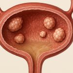
Bladder wall protrusions – often discovered incidentally during imaging for unrelated issues, or presenting as bothersome symptoms like frequent urination or discomfort – can be a source of significant concern for patients. These growths aren’t necessarily indicative of cancer, but their presence always warrants investigation to rule out malignancy and determine the best course of action. While various treatment options exist depending on the nature and size of the protrusion, endoscopic excision has emerged as a minimally invasive and effective approach for many bulky lesions. This article will delve into the specifics of this procedure, outlining what patients can expect, the potential benefits, and the considerations surrounding it.
Endoscopic excision offers an alternative to more extensive surgical interventions like partial cystectomy (bladder removal). It’s particularly well-suited for protrusions that are large enough to cause symptoms or raise suspicion but haven’t spread beyond the bladder wall. The procedure allows urologists to directly visualize the entire bladder interior, accurately assess the protrusion, and remove it with precision – all through a small incision. This results in quicker recovery times, less pain, and fewer complications compared to open surgery, making it an increasingly favored option for appropriate candidates. However, careful patient selection and thorough pre-operative evaluation are crucial for optimal outcomes.
Understanding Bulky Bladder Wall Protrusions
Bladder wall protrusions can arise from a variety of causes. These include benign conditions like inflammatory pseudotumors (inflammatory reactions mimicking tumors), diverticula (outpouchings in the bladder wall), and papillomas (wart-like growths). More seriously, they can be malignant, such as transitional cell carcinoma – the most common type of bladder cancer. Determining the exact nature of the protrusion is paramount before any treatment is considered. This typically involves a combination of imaging studies like cystoscopy (visual examination of the bladder with a camera), CT scans, and MRI. Biopsies are often taken during cystoscopy to analyze the tissue and confirm the diagnosis. To learn more about diagnostic tools, consider reviewing cystoscopic evaluations.
The size of the protrusion significantly impacts treatment decisions. Small growths may be monitored or treated with intravesical therapy (medication instilled directly into the bladder). However, bulky protrusions – generally defined as those exceeding a certain size threshold based on clinical guidelines – often require surgical intervention for definitive management. These larger lesions are more likely to cause obstructive symptoms (difficulty emptying the bladder) and pose a greater risk of being or becoming cancerous. Endoscopic excision provides a targeted way to address these growths without removing the entire bladder.
The goal of endoscopic excision isn’t simply removal; it’s achieving clear margins – meaning that all cancer cells, if present, are completely removed during the procedure. This is vital for preventing recurrence and ensuring long-term success. The technique employed must be meticulous, and adequate tissue samples should be sent to pathology for detailed analysis post-operatively. Careful follow-up monitoring is also essential after excision to detect any signs of recurrence or progression. Understanding what happens with a biopsy can give peace of mind; explore biopsy results for more information.
The Endoscopic Excision Procedure: What to Expect
The procedure itself is usually performed under general or regional anesthesia, meaning the patient will be asleep or numbed from the waist down. A cystoscope – a thin, flexible tube with a camera and light source – is inserted through the urethra (the tube that carries urine out of the body) into the bladder. The urologist then uses specialized instruments passed through the cystoscope to carefully excise the protrusion. Several techniques can be utilized for excision, including transurethral resection of bladder tumor (TURBT), which employs an electrical loop to cut and cauterize tissue, or en bloc resection, where the entire growth is removed in one piece.
Following excision, a catheter is typically left in place for several days to allow the bladder to heal and drain urine. Patients can expect some mild discomfort after the procedure, including burning during urination and blood in the urine, which usually resolves within a week or two. Most patients are able to return home the same day or the next day, depending on their overall health and the extent of the surgery. Full recovery typically takes 2-4 weeks, allowing for gradual resumption of normal activities. For those concerned about recurrence, review options for treating smaller tumors.
It’s crucial to understand that endoscopic excision doesn’t always provide a definitive cure, particularly in cases of bladder cancer. Further treatment, such as intravesical therapy (BCG or chemotherapy) or even more extensive surgery, may be necessary depending on the pathology results and the stage/grade of the tumor. A post-operative follow-up schedule will be established to monitor for recurrence and ensure optimal long-term management.
Pre-Operative Evaluation & Preparation
Before undergoing endoscopic excision, a comprehensive pre-operative evaluation is essential. This typically includes:
– A detailed medical history review, focusing on any existing health conditions and medications.
– Physical examination.
– Blood tests to assess kidney function, blood clotting ability, and overall health.
– Imaging studies (CT scan/MRI) to further evaluate the size, location, and extent of the protrusion.
– Cystoscopy with biopsy for definitive diagnosis.
Patients may be asked to discontinue certain medications, such as blood thinners, before the procedure to minimize bleeding risk. Clear instructions will be provided regarding dietary restrictions and bowel preparation (if necessary). It’s also important to discuss any allergies or sensitivities with the surgical team. This thorough preparation helps ensure a safe and successful procedure.
Intraoperative Considerations & Techniques
The success of endoscopic excision relies heavily on careful intraoperative technique. The urologist must precisely identify the boundaries of the protrusion, ensuring complete removal while minimizing damage to surrounding healthy bladder tissue. – En bloc resection is often preferred for larger or more suspicious lesions as it reduces the risk of tumor cell implantation and improves margin assessment.
During TURBT, meticulous cauterization is vital to control bleeding and achieve hemostasis. The use of advanced imaging modalities, such as narrow-band imaging (NBI), can help differentiate between cancerous and non-cancerous tissue, improving the accuracy of resection. The surgeon will also carefully assess the surrounding bladder wall for any additional lesions or areas of concern.
Post-Operative Care & Follow-Up
Post-operative care focuses on managing discomfort, preventing complications, and monitoring for recurrence. Patients should be instructed to drink plenty of fluids to flush out the urinary system and prevent clot formation. Pain medication will be prescribed as needed. Regular catheter care is essential to maintain hygiene and prevent infection.
Follow-up appointments are crucial for evaluating healing, reviewing pathology results, and discussing any further treatment options. – Cystoscopy is typically performed 3-6 months after excision to assess the bladder for recurrence. Periodic imaging studies (CT scan/MRI) may also be recommended, particularly in cases of bladder cancer. Long-term follow-up is vital for early detection of any complications or disease progression. To understand more about post op care, read through post surgical food options.


