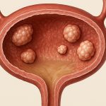
Prostatic urethral polyps (PUPs) represent a relatively uncommon but increasingly recognized entity within urological practice. Historically often misdiagnosed as benign prostatic hyperplasia (BPH) or even prostate cancer due to overlapping symptoms, advancements in diagnostic imaging and endoscopic techniques have led to improved identification and management of these lesions. Understanding PUPs is crucial for clinicians because their presence can significantly impact lower urinary tract symptoms (LUTS) and potentially mimic more serious conditions, leading to unnecessary interventions if not correctly identified. This article will delve into the nuances of endoscopic excision of prostatic urethral polyps, exploring patient selection, surgical techniques, potential complications, and post-operative management.
The diagnosis and subsequent treatment of PUPs require a careful and considered approach. While many patients may initially present with symptoms similar to BPH – including frequency, urgency, nocturia, and weak urinary stream – the underlying pathology differs considerably. The key lies in recognizing that PUPs are typically solitary lesions arising from the prostatic urethra rather than diffuse hyperplasia of the prostate gland itself. A thorough evaluation incorporating digital rectal examination (DRE), prostate-specific antigen (PSA) testing, uroflowmetry, and importantly, transrectal ultrasound (TRUS) with or without biopsy, is essential to differentiate PUPs from other conditions. Endoscopic excision offers a definitive diagnostic and therapeutic solution, allowing for histological confirmation of the polyp’s nature and immediate symptom relief.
Diagnosis and Patient Selection
Accurately diagnosing PUPs hinges on distinguishing them from other causes of LUTS. The initial clinical presentation often lacks specificity, making imaging modalities vital. TRUS plays a crucial role; while it may not always definitively identify small polyps, it can detect the presence of focal lesions within the prostatic urethra that warrant further investigation. Cystoscopy, frequently coupled with biopsy, is often the definitive diagnostic step. The endoscopic visualization allows for direct assessment of the urethral mucosa and targeted biopsies can rule out malignancy. It’s important to note that PUPs are not always associated with elevated PSA levels, although some increase may occur due to inflammation or bleeding during biopsy. To learn more about alternative approaches in similar cases, consider exploring endoscopic surgery for urethral polyps.
Patient selection for endoscopic excision is guided by several factors. Generally, patients experiencing significant LUTS disproportionate to their prostate size (suggesting a focal obstructing lesion rather than diffuse hyperplasia) and confirmed to have a urethral polyp through cystoscopic evaluation are considered candidates. The presence of a solitary polyp, readily accessible endoscopically, further supports surgical intervention. Conversely, patients with extensive or multiple polyps may require alternative management strategies, and those with significant comorbidities that increase surgical risk should be carefully evaluated before proceeding. The primary goal is to alleviate symptoms while minimizing the risk of complications. For more complex urethral issues, a surgeon might consider endoscopic resection of urethral inflammatory granulomas.
Patients presenting with suspected PUPs who demonstrate a history of recurrent urinary tract infections (UTIs), hematuria, or difficulty passing urine are also strong candidates for evaluation and potential excision. It’s critical to rule out other causes of these symptoms, such as bladder cancer or urethral stricture, before attributing them solely to a PUP. A detailed medical history, including medication use (particularly antiplatelet or anticoagulant drugs), is essential to guide pre-operative preparation and minimize bleeding risks.
Surgical Techniques for Endoscopic Excision
The gold standard treatment for symptomatic PUPs remains endoscopic excision. Several techniques can be employed, each with its advantages and disadvantages. Transurethral resection of the polyp (TURP) utilizing a loop electrode is perhaps the most commonly used method, offering precise control and effective tissue removal. However, it carries a slightly higher risk of bleeding compared to other techniques. Another approach involves using electrocautery, which provides excellent hemostasis but may be less precise for larger polyps. Holmium laser enucleation (HoLEP) is emerging as a viable option, particularly for larger or more complex PUPs, offering both precision and minimal bleeding risk.
The procedure typically follows these steps:
1. Patient positioning in the dorsal lithotomy position under spinal or general anesthesia.
2. Cystoscopic examination to confirm polyp location and size.
3. Introduction of the chosen instrument (loop electrode, electrocautery, or laser fiber) through the urethra.
4. Careful dissection around the base of the polyp, ensuring complete removal while minimizing urethral trauma.
5. Hemostasis achieved using cauterization or irrigation with epinephrine.
6. Specimen sent for histological analysis.
A critical aspect of successful excision is identifying and carefully addressing the pedicle – the stalk connecting the polyp to the urethral wall. Complete removal of the pedicle is essential to prevent recurrence. Irrigation with saline solution throughout the procedure maintains visibility and removes debris, aiding in precise dissection. The choice of technique often depends on the surgeon’s experience, the size and location of the polyp, and the patient’s overall health status. In cases requiring more specialized laser techniques, laser ablation for urethral obstruction nodules can be considered.
The use of image guidance systems during PUP excision is gaining traction. These systems enhance visualization and can improve surgical precision, particularly for smaller or more challenging polyps. Intraoperative fluoroscopy can also be utilized to confirm complete resection and guide instrument placement. Post-operatively, a Foley catheter is typically left in place for 24-72 hours to allow for urethral healing and drainage of any residual blood clots.
Potential Complications and Management
While endoscopic excision of PUPs is generally considered safe, it’s not without potential complications. The most common include postoperative bleeding, which can range from mild hematuria requiring observation to more significant bleeding necessitating transfusion or re-operation in rare cases. Urethral stricture, resulting from trauma during dissection, is another concern, although the risk is relatively low with careful surgical technique. Urinary tract infection (UTI) can occur postoperatively due to catheterization and disruption of the normal urinary flora.
Other less frequent complications include:
– Urinary incontinence – usually temporary and resolves after catheter removal.
– Retrograde ejaculation – more common with HoLEP or extensive urethral dissection.
– Urethral perforation – a rare but serious complication requiring immediate management.
Careful pre-operative evaluation to identify risk factors for bleeding (e.g., anticoagulant use) and meticulous surgical technique are paramount in minimizing complications. Postoperatively, patients should be monitored for signs of bleeding or infection. Prompt treatment with appropriate antibiotics is necessary if UTI develops. If significant bleeding occurs, cystoscopy may be required to locate the source and achieve hemostasis. In instances where urethral stricture develops, laser scalpel excision of urethral stenosis can be a viable treatment option.
Long-term follow-up is essential. Patients should undergo periodic PSA testing and cystoscopic evaluation to monitor for recurrence and assess overall urinary function. The histological analysis of the excised polyp is crucial to confirm its benign nature and guide future management decisions. If malignancy is suspected, further investigations and appropriate oncological treatment are indicated. For patients experiencing related urethral discomfort, understanding signs of urethral irritation can aid in early intervention.
Furthermore, for cases involving complex urethral issues, exploring options such as endoscopic resection of post-radiation urethral fibrosis might be necessary to address long-term complications.


