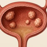
Bladder wall nodules can represent a variety of conditions, ranging from benign inflammation to more concerning neoplastic growths. Their detection often occurs during cystoscopy – a procedure where a thin, flexible tube with a camera is inserted into the bladder – and subsequent imaging. While many nodules are discovered incidentally, their presence understandably causes anxiety for patients, prompting thorough investigation and potential removal. The endoscopic approach has become increasingly sophisticated, offering minimally invasive options for both diagnosis and treatment of these lesions. It’s important to understand that removing a nodule doesn’t automatically equate to cancer; it allows for pathological examination, providing definitive answers about its nature and guiding future management strategies.
The decision to remove a bladder wall nodule is based on several factors, including the patient’s symptoms (or lack thereof), imaging characteristics of the nodule, and the individual’s overall health. Not every nodule requires removal; some can be monitored with regular cystoscopic surveillance if they appear benign. However, when there is uncertainty about the nature of a nodule or if it’s causing bothersome symptoms like frequent urination or pain, endoscopic removal is often recommended. This procedure allows for histopathological analysis, which is crucial in differentiating between inflammatory, reactive, and malignant conditions – ultimately shaping the patient’s treatment plan. The goal isn’t just to remove the tissue but to obtain a representative sample for accurate diagnosis.
Endoscopic Techniques for Nodule Removal
The core principle of endoscopic removal involves accessing the bladder through the urethra and utilizing specialized instruments to excise or vaporize the nodule while minimizing trauma to surrounding tissues. Transurethral resection of bladder tumor (TURBT) is historically the most common technique, but advancements have introduced alternatives like laser ablation and en bloc resection. TURBT utilizes an electrical loop to cut away the nodule, similar to how a polyp might be removed during a colonoscopy. While effective, it can sometimes lead to bleeding and requires careful post-operative monitoring. Laser ablation employs focused energy – typically from a holmium laser – to vaporize or coagulate the nodule. This method often results in less bleeding compared to TURBT but may not always provide sufficient tissue for comprehensive pathological examination. En bloc resection is gaining popularity, aiming to remove the entire nodule as one piece, reducing the risk of fragmentation and improving diagnostic accuracy.
The choice of technique depends on several factors including the size, location, and suspected nature of the nodule, along with the surgeon’s expertise and available equipment. Patient-specific considerations also play a role; for example, patients on blood thinners may benefit from laser ablation due to its lower bleeding risk. Importantly, all techniques require careful pre-operative assessment, including imaging studies like CT or MRI, to determine the extent of the nodule and rule out invasion beyond the bladder wall. The procedure is generally performed under spinal or general anesthesia, ensuring patient comfort during the process. Post-operatively, a catheter is typically placed for a period of time to allow the bladder to heal and prevent bleeding.
The success of any removal technique hinges on obtaining an adequate tissue sample for pathological evaluation. Fragmentation during resection can compromise diagnostic accuracy, leading to potential misdiagnosis or understaging of disease. Therefore, surgeons prioritize techniques that minimize fragmentation and ensure representative sampling. Furthermore, a thorough post-operative assessment is crucial to identify any complications like bleeding, infection, or urinary retention. Patients are typically monitored closely after the procedure and educated about warning signs to watch out for.
Complications and Management Strategies
While endoscopic removal of bladder wall nodules is generally considered safe, several potential complications can arise. Bleeding is among the most common, particularly with TURBT, but it’s usually manageable with electrocautery during the procedure or post-operative catheterization. Urinary tract infection (UTI) is another possibility, often due to instrumentation of the urethra. Strict adherence to sterile technique and prophylactic antibiotics can minimize this risk. More rarely, bladder perforation – a tear in the bladder wall – can occur, requiring surgical repair. The risk of perforation increases with larger nodules or challenging anatomical locations.
Managing post-operative complications requires prompt recognition and appropriate intervention. Significant bleeding may necessitate prolonged catheterization, blood transfusions, or even repeat endoscopic evaluation to identify and control the source of bleeding. UTIs are typically treated with antibiotics tailored to the specific organism identified in a urine culture. Bladder perforation often requires immediate surgical repair, either laparoscopically or through open surgery, depending on the severity and location of the tear. Patient education plays a vital role in minimizing complications; individuals should be informed about potential risks and warning signs to watch out for after the procedure.
A critical aspect of post-operative management is pathological analysis of the removed tissue. This determines the nature of the nodule – whether it’s benign, reactive, or malignant. If cancer is detected, further staging investigations (CT scans, MRI) are performed to determine the extent of disease and guide treatment decisions. Even if the initial nodule appears benign, ongoing surveillance with cystoscopy is often recommended to monitor for recurrence or development of new lesions. This proactive approach ensures early detection and intervention, maximizing patient outcomes.
The Role of Adjunctive Technologies
Advances in endoscopic technology are continually refining the precision and effectiveness of bladder nodule removal. Narrow band imaging (NBI) enhances visualization of subtle tissue changes, helping surgeons identify areas suspicious for cancer that might otherwise be missed. This improved visualization allows for more targeted biopsies and resection. Image-guided surgery uses real-time imaging to guide instrument placement, reducing the risk of injury to surrounding tissues and ensuring complete removal of the nodule.
Fluorescence cystoscopy, utilizing agents like hexaminolevulinate (HAL), further enhances detection capabilities. HAL is a prodrug that accumulates selectively in cancerous cells, causing them to fluoresce under blue light during cystoscopy. This allows surgeons to identify and resect areas of dysplasia or carcinoma-in-situ – early stage cancer – with greater accuracy. Robotic assistance is also emerging as a valuable tool, providing enhanced dexterity and precision for complex resections. While robotic surgery may not be suitable for all cases, it offers advantages in terms of ergonomics and visualization. Surgeons are now employing techniques like robotic-assisted excision of posterior bladder wall mass to improve outcomes.
These adjunctive technologies are not meant to replace traditional techniques but rather to complement them, improving diagnostic accuracy and surgical outcomes. They represent ongoing efforts to minimize invasiveness, enhance patient comfort, and optimize the management of bladder wall nodules. The integration of these technologies requires specialized training and expertise, ensuring that surgeons can effectively utilize their benefits while minimizing potential risks. Ultimately, a collaborative approach – combining advanced technology with skilled surgical technique – is crucial for providing optimal care to patients undergoing endoscopic removal of bladder wall nodules.


