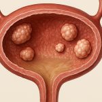
Urethral granulomas represent a relatively uncommon but often perplexing clinical challenge for urologists. These benign lesions, typically arising as a consequence of prior instrumentation, infection, or foreign body reaction, can manifest with a frustrating array of symptoms – from dysuria and frequency to more severe presentations including urethral obstruction and even acute urinary retention. Diagnosis is frequently delayed due to the non-specific nature of these complaints, often leading patients through extensive (and sometimes unnecessary) evaluations for other urological conditions. Accurate identification is paramount not just for symptomatic relief but also to differentiate granulomas from more concerning pathologies like urethral carcinoma, demanding a thoughtful and systematic approach to both diagnosis and management.
The increasing prevalence of minimally invasive urological procedures, such as transurethral resection of the prostate (TURP) and ureteroscopy, has likely contributed to an observed rise in the incidence of urethral granulomas. While many are asymptomatic and discovered incidentally during follow-up imaging, symptomatic lesions necessitate intervention to restore urinary function and improve quality of life for affected individuals. Treatment options range from conservative management with antibiotics (in suspected infectious cases) to more definitive surgical approaches, with endoscopic resection emerging as a preferred modality in many instances due to its lower morbidity compared to open surgery. This article delves into the nuances of endoscopic resection for symptomatic urethral granulomas, exploring indications, techniques, potential complications, and long-term outcomes.
Endoscopic Resection Techniques & Indications
Endoscopic resection has become a cornerstone of treatment for symptomatic urethral granulomas that are amenable to surgical intervention. The primary indication revolves around significant symptoms impacting quality of life; this could include persistent dysuria unresponsive to medical management, bothersome urinary frequency or urgency, or – most critically – evidence of urethral obstruction leading to difficulty voiding or acute retention. Preoperative evaluation typically involves cystoscopy with biopsy to confirm the granulomatous nature of the lesion and rule out malignancy. Imaging modalities such as retrograde urethrography may also be employed to delineate the extent of the granuloma and identify any associated strictures. Importantly, patients with active urinary tract infections should ideally have these resolved prior to undertaking resection to minimize postoperative complications.
The technique itself involves utilizing a resectoscope – similar to that used for TURP – but employing specialized instruments designed for urethral access. A small caliber resectoscope is generally preferred to navigate the narrower urethral channel. Resection can be performed using either monopolar or bipolar energy, with the choice often dictated by surgeon preference and available equipment. Careful dissection around the granuloma is crucial to avoid iatrogenic injury to the urethral wall. The goal isn’t necessarily complete removal of every microscopic piece of granulomatous tissue, but rather adequate resection to relieve obstruction and alleviate symptoms. Often, multiple passes are required to safely excise the bulk of the lesion.
A critical consideration in selecting patients for endoscopic resection is the location of the granuloma. Lesions located distally within the urethra – closer to the external meatus – may be more challenging to access endoscopically and might be better suited for alternative approaches, such as direct excision under local anesthesia. Conversely, proximal lesions are typically easier to visualize and resect with standard cystoscopic techniques. Furthermore, patients with significant urethral strictures accompanying the granuloma may require prior or concurrent urethroplasty to optimize long-term outcomes.
Postoperative Management & Complications
Following endoscopic resection, a temporary catheter is usually placed for several days to allow for healing and prevent edema from causing obstruction. The duration of catheterization varies based on the size and location of the granuloma as well as individual patient factors. Patients are typically instructed to drink plenty of fluids postoperatively to promote urinary flow and minimize the risk of clot formation. Close monitoring for signs of infection or bleeding is essential during this period. Antibiotics may be prescribed prophylactically, especially if there’s concern for underlying infection contributing to the granuloma’s development.
Several potential complications can arise following endoscopic resection. – Urethral stricture remains one of the most significant long-term concerns, as the surgical trauma itself can contribute to scar tissue formation and narrowing of the urethral channel. This risk is heightened in patients with pre-existing mild strictures. – Bleeding is another potential complication, although typically minor and controlled during the procedure. Significant bleeding requiring transfusion is rare but possible. – Urethral perforation represents a more serious, though uncommon, complication that necessitates immediate recognition and management.
Preventing complications involves meticulous surgical technique, careful patient selection, and appropriate postoperative care. Patients should be counseled about the risk of stricture formation and the importance of follow-up cystoscopy to monitor for recurrence or narrowing of the urethra. In cases where stricture develops, subsequent dilation or urethroplasty may be necessary to restore urinary flow.
Recurrence & Long-Term Outcomes
Unfortunately, despite successful initial resection, recurrence rates for urethral granulomas can be relatively high – ranging from 20% to 40% in some studies. This is likely due to the underlying etiology of the lesion; if the inciting factor (e.g., chronic irritation from a foreign body or persistent low-grade infection) isn’t addressed, the granuloma may reform over time. Therefore, identifying and mitigating these contributing factors is crucial for minimizing recurrence. For example, addressing any residual fragments of prostatic calculi after TURP or managing underlying bladder dysfunction can help reduce the likelihood of repeat granuloma formation.
Long-term outcomes following endoscopic resection are generally favorable for patients experiencing symptomatic relief. Most individuals report a significant improvement in urinary symptoms and quality of life. However, ongoing surveillance is recommended to detect recurrence or development of complications such as urethral strictures. Follow-up cystoscopy should be performed at regular intervals – typically every 6 to 12 months – for the first few years after resection, then less frequently if no issues arise.
The need for repeat endoscopic procedures varies depending on individual patient factors and the location/size of recurrent granulomas. In some cases, alternative treatment strategies such as direct excision or even conservative management with intermittent catheterization may be considered if further resection is deemed undesirable or unlikely to provide lasting benefit. The overall success of endoscopic resection hinges on a comprehensive approach that encompasses accurate diagnosis, meticulous surgical technique, appropriate postoperative care, and diligent long-term follow-up.


