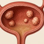
Radiation cystitis represents a significant morbidity for patients undergoing radiotherapy for pelvic malignancies, including prostate, cervical, and rectal cancers. The inflammation induced by radiation damages the bladder mucosa, leading to fibrosis – essentially scarring – within the bladder wall. This fibrosis reduces bladder capacity, increases urinary frequency and urgency, and can cause debilitating pain. While conservative management options exist, including medications to manage symptoms and hyperbaric oxygen therapy in select cases, significant fibrous lesions often require surgical intervention. The goal of surgery isn’t necessarily to cure radiation cystitis – the underlying damage remains – but rather to improve bladder function and quality of life by addressing the most problematic areas of fibrosis. Successful management hinges on careful patient selection, meticulous surgical technique, and a comprehensive understanding of the challenges posed by irradiated tissues.
The development of these fibrous lesions is a progressive process often taking months or even years after radiation therapy completion. Initial inflammation gives way to chronic tissue damage and eventually, significant fibrotic changes. These lesions can manifest in various forms – from diffuse thickening of the bladder wall to discrete, localized masses. Importantly, differentiating between radiation-induced fibrosis and tumor recurrence is paramount before considering surgical intervention; imaging studies (CT, MRI) and cystoscopy with biopsies are essential for accurate diagnosis. The decision to proceed with excision is not taken lightly, as surgery in irradiated tissues carries inherent risks including increased bleeding potential, wound healing complications, and the possibility of creating further bladder damage. However, for patients experiencing substantial functional impairment or intractable pain, surgical intervention can offer a path towards improved quality of life.
Surgical Approaches to Fibrous Bladder Lesion Excision
The optimal surgical approach to excising fibrous lesions in radiation cystitis is highly individualized, dependent on the location, size, and number of lesions, as well as the overall condition of the patient. Open surgery (laparotomy) has historically been the standard, allowing for thorough exploration of the bladder and surrounding structures. However, robotic-assisted laparoscopic surgery (RALS) is increasingly utilized, offering advantages such as enhanced visualization, improved dexterity, and potentially faster recovery times. A transurethral approach – using cystoscopic instruments passed through the urethra – can be considered for smaller, accessible lesions but is limited by its ability to address extensive fibrosis or deeply situated masses. Choosing the right technique requires a careful assessment of each patient’s specific needs and surgical expertise available.
The fundamental principle behind all excision techniques is to remove the fibrotic tissue while preserving as much functional bladder wall as possible. During open surgery or RALS, surgeons will carefully dissect around the lesion, utilizing electrocautery and meticulous hemostasis (bleeding control) due to the fragility of irradiated tissues. A key consideration is avoiding injury to surrounding structures like the ureters and rectum. After excision, the resulting defect in the bladder wall may be closed primarily if small enough, or reconstructed using a flap of healthy bladder tissue or even a segment of bowel – although this is reserved for more extensive resections. The goal is always to maintain adequate bladder capacity and prevent future complications such as fistula formation.
The selection of patients who will benefit from surgical excision requires stringent criteria. Patients with significant co-morbidities, those with widespread diffuse fibrosis affecting the entire bladder, or those without a clear diagnosis (where tumor recurrence cannot be ruled out) are generally not considered good candidates. Preoperative assessment includes detailed imaging to delineate the extent of fibrosis, urodynamic studies to assess bladder function, and thorough evaluation for any contraindications to surgery. Patient counseling is crucial – outlining the potential benefits, risks, and alternatives to surgical intervention is essential for informed consent.
Considerations During Surgery
The irradiated nature of the tissues presents unique challenges during surgery. – Increased Bleeding: Irradiated tissues are more prone to bleeding due to damage to blood vessels and impaired coagulation factors. Meticulous hemostasis throughout the procedure is vital, often requiring careful suture placement and potentially the use of topical hemostatic agents.
– Impaired Wound Healing: Radiation significantly compromises wound healing capacity. This increases the risk of postoperative complications such as wound dehiscence (separation) and infection. Prophylactic antibiotics are routinely used, and surgeons take extra care to minimize tissue trauma during dissection.
– Tissue Friability: The fibrotic tissues can be fragile and easily torn, making dissection more difficult and increasing the risk of inadvertent injury to surrounding structures. Gentle handling and precise surgical technique are paramount.
Surgeons must also be vigilant for signs of ongoing inflammation or infection within the bladder. Preoperative cultures may be helpful in identifying any existing bacterial colonization. During surgery, irrigation with antibiotic solutions can further reduce the risk of postoperative infections. The use of minimally invasive techniques (RALS) can potentially minimize tissue trauma and improve wound healing compared to open surgery.
Postoperative Management & Potential Complications
Postoperative care following excision of fibrous bladder lesions in radiation cystitis is critical for optimizing outcomes and preventing complications. Patients require close monitoring for signs of infection, bleeding, and urinary leakage. A Foley catheter is typically left in place for several weeks to allow the surgical site to heal and prevent urine extravasation (leakage). Regular follow-up appointments are essential to assess bladder function, monitor for recurrence of symptoms, and address any complications that may arise.
Potential postoperative complications include: – Urinary Fistula: An abnormal connection between the bladder and another organ or skin surface, leading to continuous urine leakage.
– Bladder Perforation: Accidental puncture of the bladder wall during surgery, requiring repair.
– Wound Infection: Increased risk due to impaired wound healing in irradiated tissues.
– Recurrence of Fibrosis: Despite successful excision, fibrosis can recur over time, necessitating further intervention.
– Urinary Retention: Inability to empty the bladder fully, potentially requiring intermittent catheterization.
Long-term management often involves ongoing urodynamic monitoring and symptomatic treatment. Patients may require continued medications to manage urinary symptoms or undergo additional procedures such as bladder augmentation (increasing bladder capacity) if necessary. The success of surgical intervention is not solely determined by the immediate outcome but also by the ability to provide comprehensive long-term care and support to these patients.
The Role of Urodynamic Studies & Future Directions
Urodynamic studies are essential both preoperatively and postoperatively in managing radiation cystitis. Preoperative studies help assess baseline bladder capacity, compliance (how well it stretches), and flow rates, providing valuable information for surgical planning and patient selection. Postoperative studies monitor changes in bladder function after excision of fibrous lesions, helping to identify any complications or recurrence of symptoms. Specifically, they can detect decreased bladder capacity, impaired detrusor function (the muscle responsible for bladder emptying), and urinary leakage.
Looking ahead, research is focused on developing strategies to prevent radiation cystitis before it develops. This includes exploring the use of radioprotective agents during radiotherapy and optimizing radiation techniques to minimize collateral damage to the bladder. New surgical approaches are also being investigated, such as robotic-assisted interstitial ablation (using heat to destroy fibrotic tissue) and gene therapy aimed at promoting tissue regeneration. Ultimately, a multidisciplinary approach involving radiation oncologists, urologists, and reconstructive surgeons is crucial for providing optimal care to patients with radiation cystitis and maximizing their quality of life. The goal remains to alleviate the debilitating symptoms associated with this condition and improve long-term bladder function.


