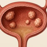
Bladder cancer represents a significant global health concern, impacting hundreds of thousands of individuals annually. Early detection and accurate staging are paramount for effective treatment and improved patient outcomes. Traditionally, cystoscopy – the visual examination of the bladder using a flexible scope – has been the cornerstone of diagnosis and management. However, relying solely on white light cystoscopy can be limited, often missing small or flat lesions that may harbor cancerous cells. This is where high-definition (HD) cystoscopy-assisted bladder tumor mapping emerges as a critical advancement, offering enhanced visualization and precision in identifying and characterizing these potentially life-threatening growths. It’s not simply about seeing more; it’s about seeing better and with greater certainty.
The limitations of conventional cystoscopy have spurred the development of technologies aimed at improving detection rates and reducing recurrence. HD cystoscopy builds upon this foundation, employing higher resolution imaging to reveal subtle morphological changes within the bladder wall that might be missed by standard techniques. Furthermore, adjunctive imaging modalities – like narrow band imaging (NBI) or image-guided fluorescence (IGF) using agents like pentinoxime green (PGI) – are frequently integrated into HD cystoscopy protocols to highlight cancerous tissue and differentiate it from healthy mucosa. Tumor mapping then takes this enhanced visualization a step further by systematically documenting the location, size, and characteristics of all detected lesions, creating a comprehensive “map” of the bladder’s interior for treatment planning and follow-up monitoring. This approach is becoming increasingly vital in optimizing patient care pathways.
The Role of High-Definition Cystoscopy & Adjunctive Imaging
HD cystoscopy itself represents a substantial leap forward from older technologies. The increased pixel density provides sharper, more detailed images, allowing urologists to discern subtle differences in tissue texture and vascular patterns that could indicate malignancy. This improved resolution is particularly valuable when evaluating patients with non-muscle invasive bladder cancer (NMIBC), where early detection of in situ carcinoma or low-grade tumors can significantly impact treatment decisions. However, HD alone isn’t always enough. Adjunctive imaging techniques are often used to augment the diagnostic accuracy and sensitivity of cystoscopy.
Narrow band imaging utilizes specific wavelengths of light that enhance visualization of blood vessels and mucosal surface features. Cancerous tissue typically exhibits altered vascularity – displaying irregular or dilated vessels – making it more readily identifiable against a background of normal mucosa. Image-guided fluorescence, utilizing agents like PGI which selectively adhere to cancerous cells, further enhances lesion detection by causing them to fluoresce under blue light. The combination of HD cystoscopy with these adjunctive modalities dramatically improves the ability to identify lesions that would otherwise be missed, leading to more accurate staging and potentially reducing the risk of recurrence. This multi-faceted approach is becoming standard practice in many comprehensive cancer centers. Understanding bladder tumor staging helps determine the best course of action.
The selection of an appropriate imaging modality often depends on factors like patient history, tumor grade, and institutional protocols. Some institutions prioritize NBI for routine cystoscopy, while others favor IGF with PGI for patients at higher risk of recurrent disease. The key takeaway is that integrating these technologies alongside HD cystoscopy significantly improves diagnostic capabilities compared to white light cystoscopy alone.
Bladder Tumor Mapping: A Systematic Approach
Bladder tumor mapping isn’t simply about finding lesions; it’s a rigorous, systematic process designed to create a complete and accurate record of all findings. This meticulous documentation is critical for several reasons – including guiding transurethral resection of bladder tumors (TURBT), planning subsequent surveillance cystoscopies, and comparing results over time to assess treatment response and detect recurrence. A well-executed mapping procedure ensures that no lesions are overlooked and provides a baseline for future monitoring.
The process typically involves a standardized approach to examining the entire bladder surface in a methodical manner. This often begins with visualizing the urethra and then proceeding systematically around the bladder, documenting each lesion encountered. Information recorded during mapping includes: – Location (using anatomical landmarks or clock-face descriptions) – Size (measured in millimeters) – Morphology (e.g., papillary, flat, ulcerated) – Grade (based on histological assessment of biopsy samples) – This often requires taking multiple biopsies from each lesion and surrounding areas.
The accuracy of bladder tumor mapping is heavily reliant on the skill and experience of the urologist performing the procedure, as well as the quality of the imaging equipment used. Increasingly, digital cystoscopy systems are being adopted to facilitate more precise documentation and image archiving. These systems allow for real-time recording of high-resolution images and videos, which can be easily reviewed and shared among healthcare professionals. This technology is proving invaluable in improving communication and collaboration within multidisciplinary teams dedicated to bladder cancer care. Surgeons may also consider robotic assisted resection for increased precision.
Optimizing TURBT with Mapping Data
Transurethral resection of bladder tumor (TURBT) is often the primary treatment for NMIBC. The goal of TURBT is to completely remove all visible tumors while minimizing damage to healthy tissue. However, incomplete resection can significantly increase the risk of recurrence. This is where accurate bladder tumor mapping plays a crucial role in optimizing the surgical procedure.
By providing a detailed roadmap of lesion locations and characteristics, tumor mapping allows the surgeon to precisely target areas requiring resection. It helps avoid unnecessary removal of healthy mucosa, reducing postoperative complications like bleeding and perforation. Furthermore, it ensures that all visible lesions are addressed during the initial surgery, maximizing the chances of achieving complete tumor eradication. Some surgeons now use real-time intraoperative imaging – often coupled with fluorescence guidance – to further refine their resection technique based on the mapping data. The integration of mapping data into TURBT significantly improves surgical precision and completeness. A TURBT procedure is often the first step in treatment.
Surveillance Cystoscopies & Recurrence Monitoring
Even after successful TURBT, patients with NMIBC require regular surveillance cystoscopies to monitor for recurrence. These follow-up examinations are essential because bladder cancer has a relatively high rate of recurrence, even in low-risk tumors. Bladder tumor mapping is invaluable during these surveillance visits, providing a baseline against which to compare subsequent findings.
By comparing the location and characteristics of new lesions with those documented in previous mappings, urologists can more accurately assess disease progression and identify areas of concern. This allows for timely intervention – such as repeat TURBT or adjunctive therapies – if recurrence is detected. The use of digital cystoscopy systems facilitates this comparison by providing easy access to archived images from prior examinations. The ability to track changes over time provides a much clearer picture of the disease course and helps guide treatment decisions.
Future Directions & Technological Advancements
The field of HD cystoscopy-assisted bladder tumor mapping is constantly evolving, with ongoing research focused on further enhancing detection accuracy and improving patient outcomes. Several promising technologies are currently under development that have the potential to revolutionize this area.
One key area of innovation involves artificial intelligence (AI) and machine learning algorithms. These AI systems can be trained to analyze cystoscopic images and automatically identify suspicious lesions, potentially assisting urologists in real-time during examinations. Another exciting development is the use of advanced imaging modalities like photoacoustic tomography (PAT), which combines optical and ultrasound techniques to provide high-resolution visualization of both superficial and deeper bladder tissues. Finally, improvements in image-guided drug delivery systems are being explored to allow for targeted therapy directly to cancerous lesions identified during cystoscopy. These advancements promise a future where bladder cancer detection and treatment are even more precise, personalized, and effective. Understanding recurrence risk is crucial for long term patient care, as well as the impact of a recurrence on symptoms.


