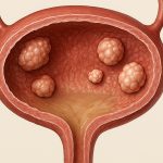
Posterior bladder wall masses represent a diagnostic and surgical challenge for urologists. These lesions, often discovered incidentally during imaging for unrelated issues or presenting as hematuria (blood in the urine) or altered voiding habits, demand careful evaluation to differentiate between benign growths like fibromas or inflammatory processes and malignant tumors such as urothelial carcinoma – bladder cancer. The location itself presents difficulties; the posterior wall is less accessible than other parts of the bladder, making traditional open surgery more invasive and potentially impacting patient recovery. Historically, transurethral resection (TUR) was often employed, but its effectiveness can be limited depending on the size, depth, and characteristics of the mass.
The advent of robotic-assisted surgery has provided a compelling alternative to both open and conventional laparoscopic approaches for excising these posterior bladder wall masses. Robotic platforms offer enhanced precision, improved visualization via three-dimensional imaging, and greater dexterity through instruments with seven degrees of freedom – mimicking human hand movements more accurately than traditional laparoscopic tools. This translates into potentially less blood loss, reduced postoperative pain, shorter hospital stays, and faster return to normal activities for patients undergoing this procedure. Importantly, robotic assistance doesn’t replace the surgeon; it augments their skills, allowing for a more refined surgical technique in a challenging anatomical location. Surgeons can now utilize techniques like robotic mass mapping and resection to precisely target the affected area.
Preoperative Evaluation and Patient Selection
A thorough preoperative evaluation is absolutely critical before considering robotic-assisted excision of a posterior bladder wall mass. This begins with detailed imaging studies to characterize the lesion accurately. – Cystoscopy, both white light and narrow band imaging (NBI), provides direct visualization of the bladder interior and helps assess the size, location, and appearance of the mass. – Computed tomography (CT) scans or magnetic resonance imaging (MRI) are essential for staging purposes, evaluating potential spread beyond the bladder wall (T-staging), and identifying any involvement of surrounding structures like the rectum or pelvic lymph nodes. Importantly, MRI is often preferred as it provides superior soft tissue resolution compared to CT. – Urine cytology, which examines cells shed from the bladder lining, can help detect evidence of malignancy even if cystoscopy appears benign.
Patient selection is equally important. Robotic-assisted excision isn’t appropriate for all patients with posterior bladder wall masses. Ideal candidates are generally those with: – Non-muscle invasive bladder cancer (NMIBC) confined to the posterior wall – robotic assistance allows for precise resection while minimizing the risk of damaging healthy tissue. – Masses that are accessible robotically, meaning they aren’t too large or situated in a location where robotic instruments can’t effectively reach them. – Patients who are medically fit enough to undergo general anesthesia and robotic surgery. Those with significant comorbidities (other health conditions) may be better suited for alternative approaches. Careful consideration of patient factors is paramount. Understanding tumor invasion patterns helps guide these decisions.
The evaluation process also includes a detailed discussion with the patient about the risks and benefits of robotic-assisted excision compared to other treatment options, such as TURBT (transurethral resection of bladder tumor), open surgery or observation if appropriate. Patient expectations should be managed realistically, emphasizing that while robotic assistance offers several advantages, it is not without potential complications.
Surgical Technique: A Step-by-Step Overview
The robotic-assisted excision typically involves a minimally invasive approach utilizing small incisions through which the robotic arms and camera are inserted. The procedure generally follows these steps: 1. Patient Positioning: The patient is positioned in lithotomy (legs spread) to provide optimal access to the bladder. Pneumoperitoneum is established – inflating the abdomen with carbon dioxide gas to create space for surgical visualization and manipulation. 2. Docking the Robot: The Da Vinci Surgical System is then docked, aligning the robotic arms over the patient’s lower abdomen. The surgeon operates from a console, controlling the robot’s movements with hand and foot controls. 3. Bladder Mobilization: Gentle dissection around the bladder is performed to mobilize it and create a clear surgical field. This often involves identifying key anatomical landmarks like the ureters (tubes carrying urine from kidneys to bladder) and rectus abdominis muscles. 4. Mass Resection: The posterior wall mass is carefully resected using robotic instruments, ensuring adequate margins – removing surrounding tissue to minimize the risk of recurrence. The extent of resection depends on the pathological findings after initial biopsy or TURBT. This step requires precise technique and visualization. 5. Bladder Reconstruction: After mass removal, any defects in the bladder wall are typically reconstructed using absorbable sutures, creating a smooth and watertight closure. The goal is to restore normal bladder function as much as possible. 6. Drainage & Closure: A small drain may be placed near the bladder to help remove any postoperative fluids. The abdominal incisions are then closed with sutures or staples.
Throughout the procedure, continuous monitoring of vital signs and careful attention to hemostasis (stopping bleeding) are crucial. The surgeon constantly assesses the surgical field through the 3D robotic vision system, making adjustments as needed to ensure optimal outcomes. Meticulous technique is key. Consideration should be given to bladder mass excision with closure during the reconstruction phase.
Intraoperative Considerations & Avoiding Complications
Several intraoperative considerations are paramount for a successful robotic-assisted excision. One of the most significant concerns is ureteral injury – accidental damage to the tubes carrying urine from the kidneys. This can be avoided by carefully identifying and protecting the ureters during dissection and resection. Utilizing techniques like ureteral stents (small tubes inserted into the ureter) preoperatively can further aid in visualization and protection. Another potential complication is bladder perforation, which can occur during dissection or mass removal. If a perforation occurs, it must be promptly identified and repaired robotically or converted to an open approach if necessary.
Maintaining adequate hemostasis – controlling bleeding – is also critical. Robotic instruments allow for precise coagulation (sealing of blood vessels) using energy modalities like bipolar or harmonic scalpel. – Frequent irrigation of the surgical field helps improve visualization and remove any blood clots. – The use of a dedicated suction device is essential to maintain a dry operative field. Proactive management of bleeding minimizes postoperative complications.
Finally, careful attention must be paid to preserving bladder function. Excessive resection can lead to detrusor instability (involuntary bladder contractions) or urge incontinence. The goal is always to remove the mass completely while minimizing damage to surrounding healthy tissue and preserving as much functional bladder capacity as possible. Techniques for bladder wall flap closure are often utilized during this phase.
Postoperative Recovery & Long-Term Follow-up
Postoperative recovery following robotic-assisted excision of a posterior bladder wall mass is generally faster and less painful compared to open surgery. Patients typically experience: – Shorter hospital stays – often discharged within 1-3 days. – Reduced postoperative pain, managed with oral analgesics (pain medication). – Faster return to normal activities, usually within several weeks. A Foley catheter (tube inserted into the bladder) is generally left in place for a few days to allow the bladder to heal and prevent leakage. Patients are instructed on proper catheter care and monitoring for signs of infection.
Long-term follow-up is crucial to monitor for recurrence or progression of disease. This typically involves: – Regular cystoscopies – every 3-6 months initially, then annually – to examine the bladder lining for any new tumors. – Urine cytology – performed periodically to detect recurrent cancer cells in the urine. – Imaging studies (CT/MRI) – as needed based on clinical findings. Consistent follow-up is essential for early detection and management of recurrence. Patients are also educated about signs and symptoms of bladder cancer, such as hematuria or changes in voiding habits, and encouraged to report any concerns promptly. For patients with more complex cases, robotic assistance can extend beyond the bladder.


